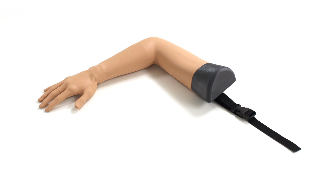

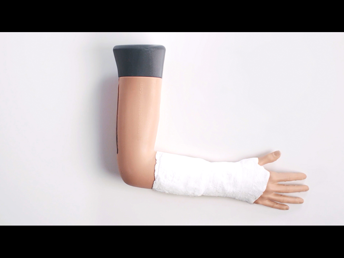
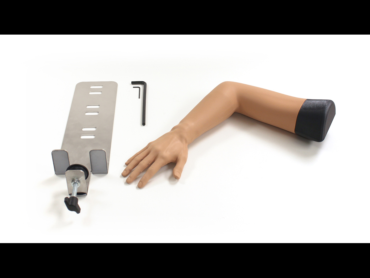
Colles' Fracture Reduction Trainer (Light Skin Tone)
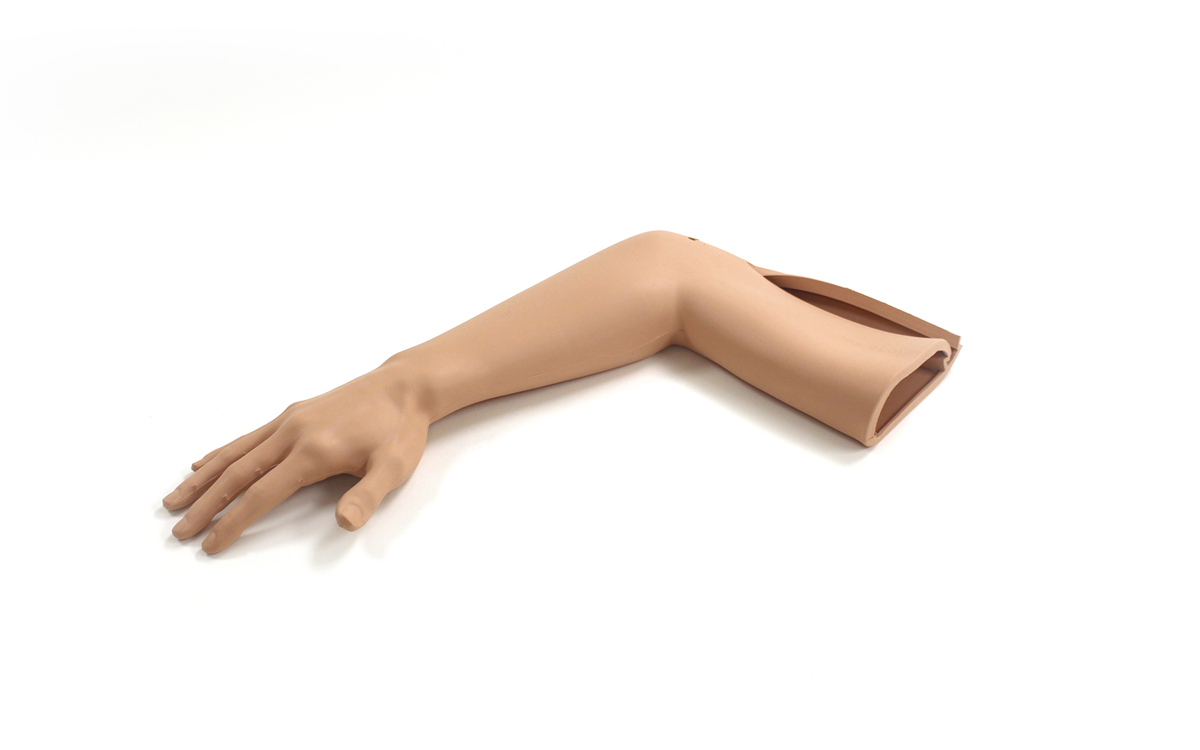
Designed for Emergency Medicine and Orthopaedic Specialist trainees, the Limbs & Things Colles’ Fracture Reduction Trainer offers repeatable practice in fracture reduction of the wrist. It is also suitable for training junior doctors, Emergency Nurse Practitioners, staff nurses, Orthopaedic Practitioners (or Plasterer Technicians) and Health Care Assistants (HCAs).
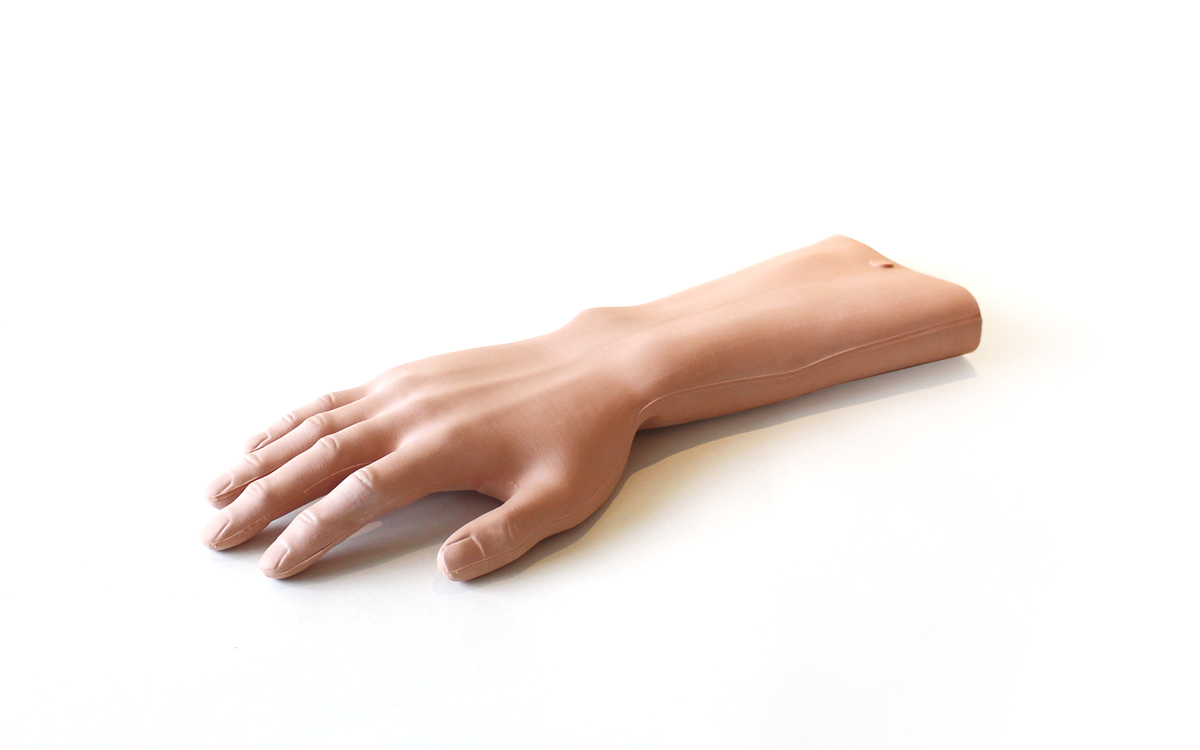
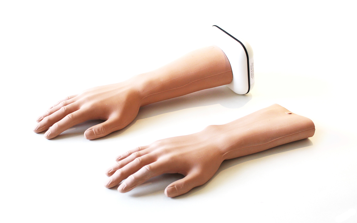
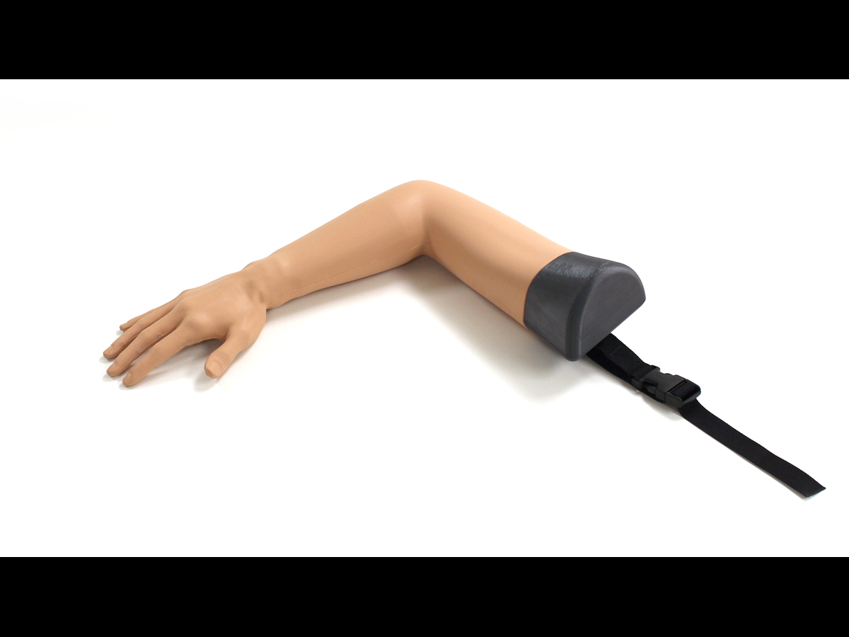
The Colles’ Fracture Reduction Trainer represents a fractured distal fragment of the radius and enables trainees to carry out the full procedure, diagnosis through visual inspections, reduction procedure and plastering the arm.
For more accurate real-life simulations, the Colles’ trainer has an adjustable tension mechanism that allows for various levels of difficulty, making it ideal for advanced learning. The design also allows for common mistakes to occur such as a partially reduced fractures, and if the arm is not held correctly when plastered, it will pop back to the original state.
The model has been designed in collaboration with Dr Salwa Malik of Brighton and Sussex University Hospitals NHS Trust and Dr Yasir M. Shaukat of Surrey and Sussex Hospitals NHS Trust.
Overview
- Adjustable tension allows for progressive levels of difficulty
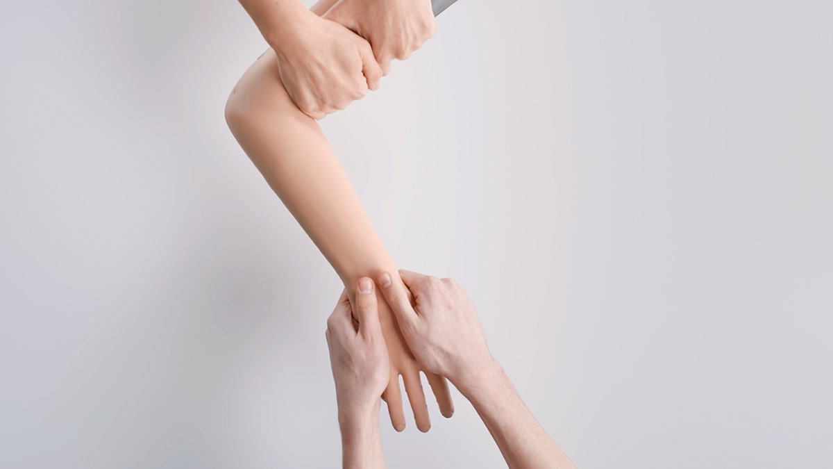
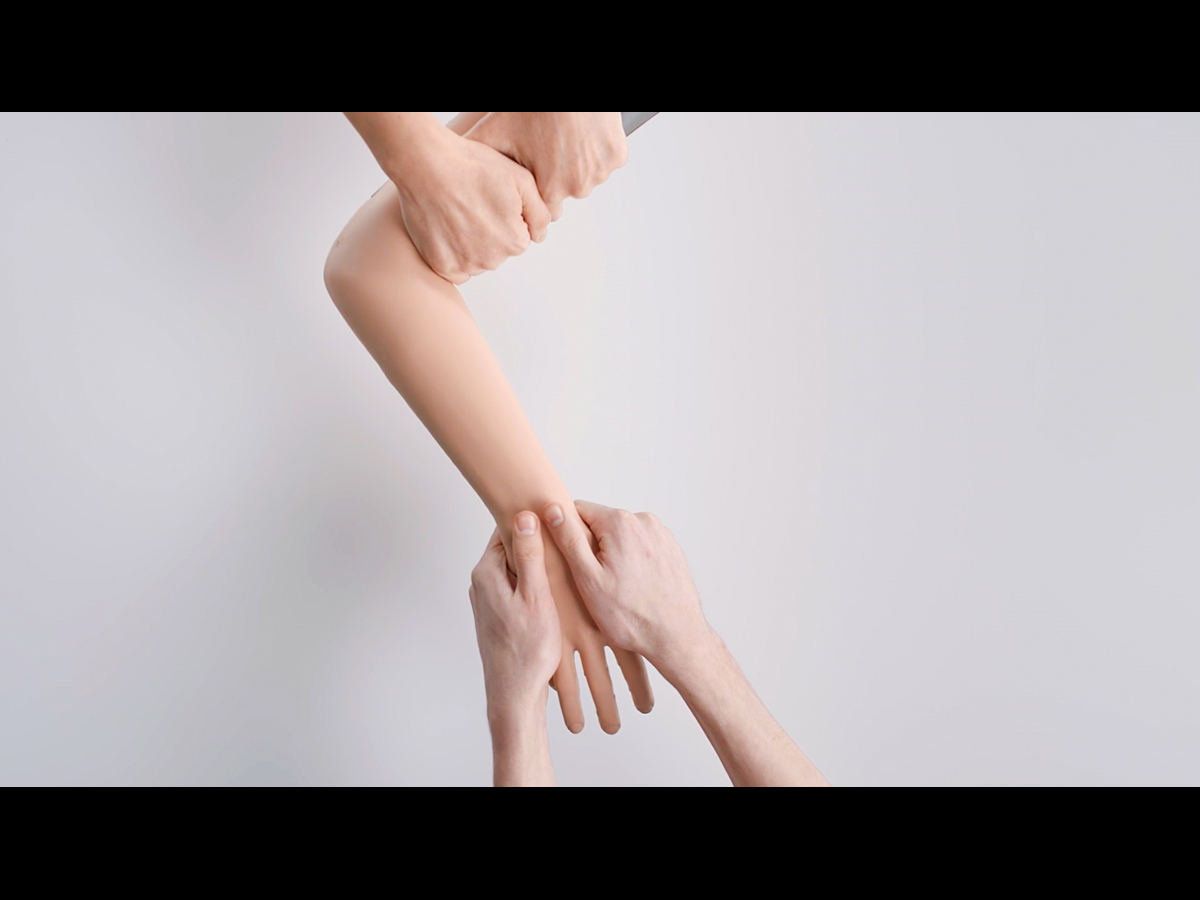
- Trainees can perform all three stages of reduction: Exaggeration (to dis-impact the fracture), Traction and Flexion
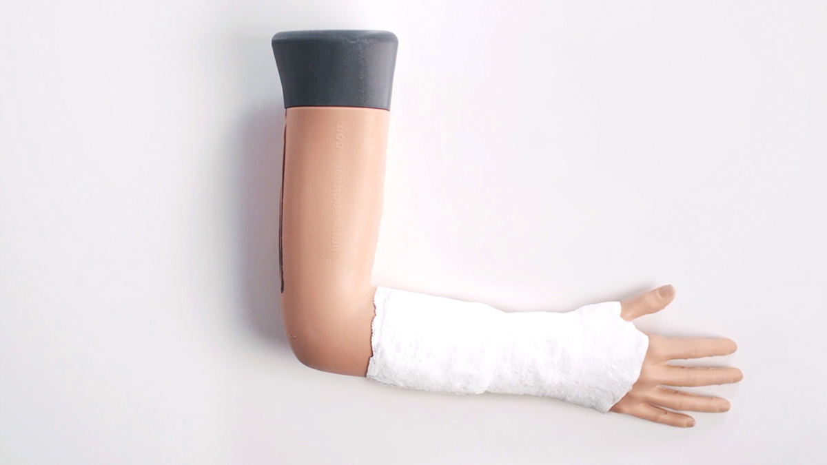
- Accommodates various plastering techniques, including back slab, circular cast, sugar-tong splint, and 3-point moulds
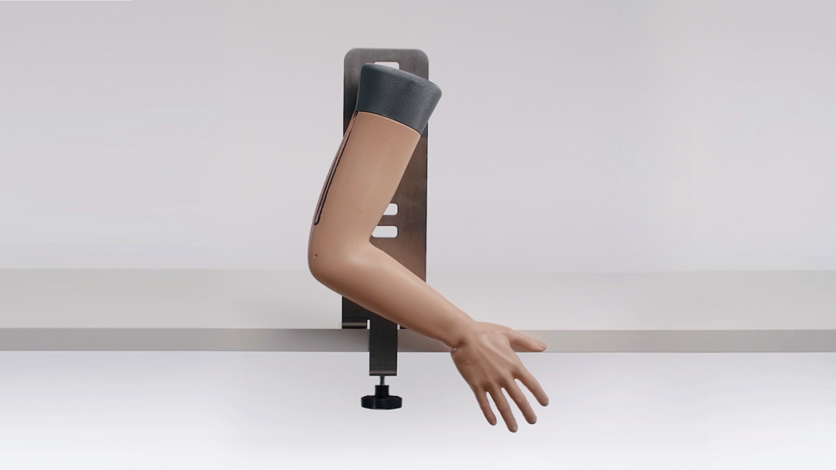
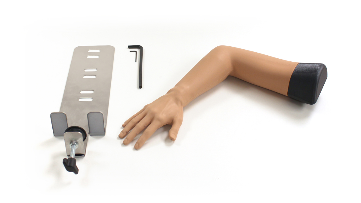
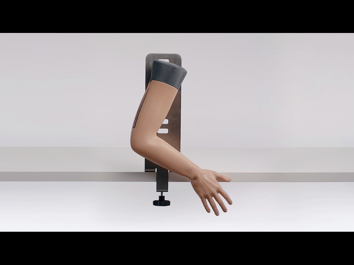
- Stand allows the Colles’ Fracture arm to be placed at a variety of heights for different patient scenarios
Realism
- Fractured distal fragment of radius, and head & body of the ulna bone for visual and palpable identification
- Realistic representation of a distal angulation (dinner-fork) deformity
- Life-like haptic feedback when conducting the procedure
Versatility
- Adjustable tension makes this trainer a perfect tool for OSCE courses
- The stand allows the Colles’ Fracture arm to be placed at a variety of heights for different patient scenarios
Cleaning
- Clean skin surface with soft, damp cloth and mild detergent
Safety
- This product is latex free
Simulated Patient
- This product can be used with a simulated patient
Anatomy
- Fractured distal fragment of radius
- Head and body of the ulna
Skills Gained
- Visual inspection & identification
- Reduction of the fracture including Exaggeration (to dis-impact the fracture), Traction and Flexion
- Plastering techniques
- Teamwork and communication when reducing the fracture
References
Accreditation Council for Graduate Medical Education. Closed treatment of distal radial fracture (e.g., Colles or Smith type) or epiphyseal separation, includes closed treatment of fracture of ulnar styloid when performed; with manipulation
Accreditation Council for Graduate Medical Education. Closed treatment of distal radial fracture (e.g., Colles or Smith type) or epiphyseal separation, includes closed treatment of fracture of ulnar styloid when performed; without manipulation
Accreditation Council for Graduate Medical Education. Demonstrates knowledge of associated hand injury patterns (e.g., median nerve injury and/orscapholunate ligament (SL) injury with distal radius fractures); Performs simple closed reduction and splinting of pediatric and adult fractures/dislocations
American Academy of Family Physicians. In the absence of significant displacement, comminution, or intra-articular involvement, many forearm fractures can be managed by a primary care physician. Initial management of forearm fractures should follow the PRICE (protection, rest, ice, compression, and elevation) protocol, except forcompression, which should be avoided in the acute setting. Distal radius fractures with minimal displacement can be treated with a short arm cast
Orthopedic Injuries, Clerkship Directors in Emergency Medicine. Reduction of the fracture or dislocation–for closed fractures or dislocations, reduction takes precedence when there is any limb ischemia or lack of pulses and emergent reduction is required. It is important to reduce a fracture or dislocation to restore anatomic appearance, alleviate pain, and relieve tension on the tissues.This most often requires appropriate sedation and monitoring of the patient and can be done at the bedside in the ED with procedural sedation.After a reduction has been performed, obtain a post-reduction imaging to ensure successful alignment.
Curriculum for Education and Training in Orthopaedic Surgery: Overview, Australian Orthopaedic Association, 2017. SECTION 3 – APPLIED MEDICAL AND SURGICAL EXPERTISE IN ORTHOPAEDICS 3.4 Hand and Wrist ME - Applied Sciences Anatomy, including surgical approaches Biomechanics Pathology ME - Assessment History taking Physical Examination Investigations
Emergency Medicine Certificate and Diploma, Australasian College for Emergency Medicine, 2019. Undertake history, examination, investigation and initiate treatment of patients presenting with orthopaedic trauma Safely and appropriately carry out the following procedures: Simple joint reductions,Interpretation of plain radiology, Application of plaster-of paris backslab to forearm and lower limb including appropriate aftercare, Application of digital nerve block
Curriculum and Assessment Systems for Core Specialty Training ACCS CT1-3 & Higher Specialty Training ST4-6 Training Programmes, The College of Emergency Medicine, p. 245 and p.344, 2012).
Trauma & Orthpaedic Surgery Curriculum, The Intercollegiate Surgical Curriclum Programme, 2021, p.171.
Can a single person perform training with the Colles’ distal radius model?
The Colles trainer should always be used with a minimum of two people, conducting traction and counter traction, even when using the stand.
What are the Hex Keys used for that are supplied with the Colles’ Fracture Reduction Trainer?
The Hex Keys are used to adjust the tension mechanism, allowing trainers to alter the difficulty of the procedure for advanced learning.
What size are the Hex Keys used with the Colles’ Fracture Trainer?
The Larger Hex Key is 5mm and the Smaller Hex Key is 2.5mm.
The Imperial equivalent is 3/16 for the larger Hex and 3/32 for the smaller one.
Does the Colles’ Fracture Reduction Trainer allow for visualisation through ultrasound?
This trainer has been designed for physical assessment and Colles’ fracture reduction procedures and doesn’t possess any ultrasound capabilities.

