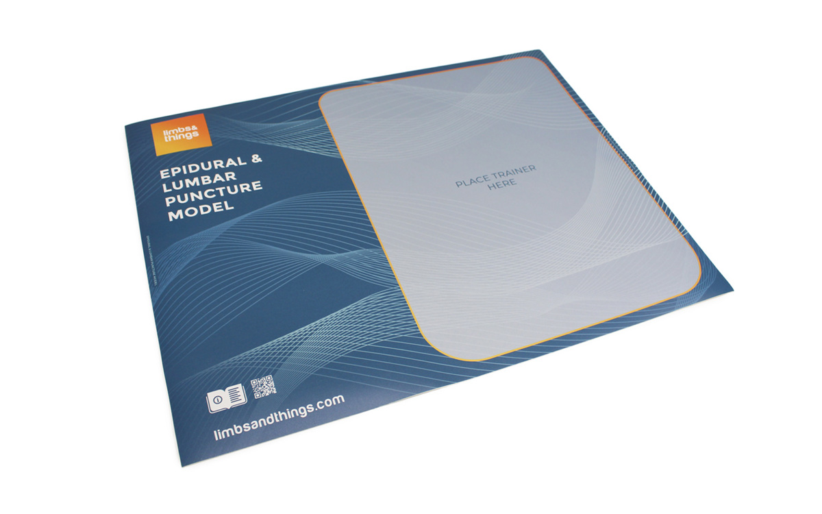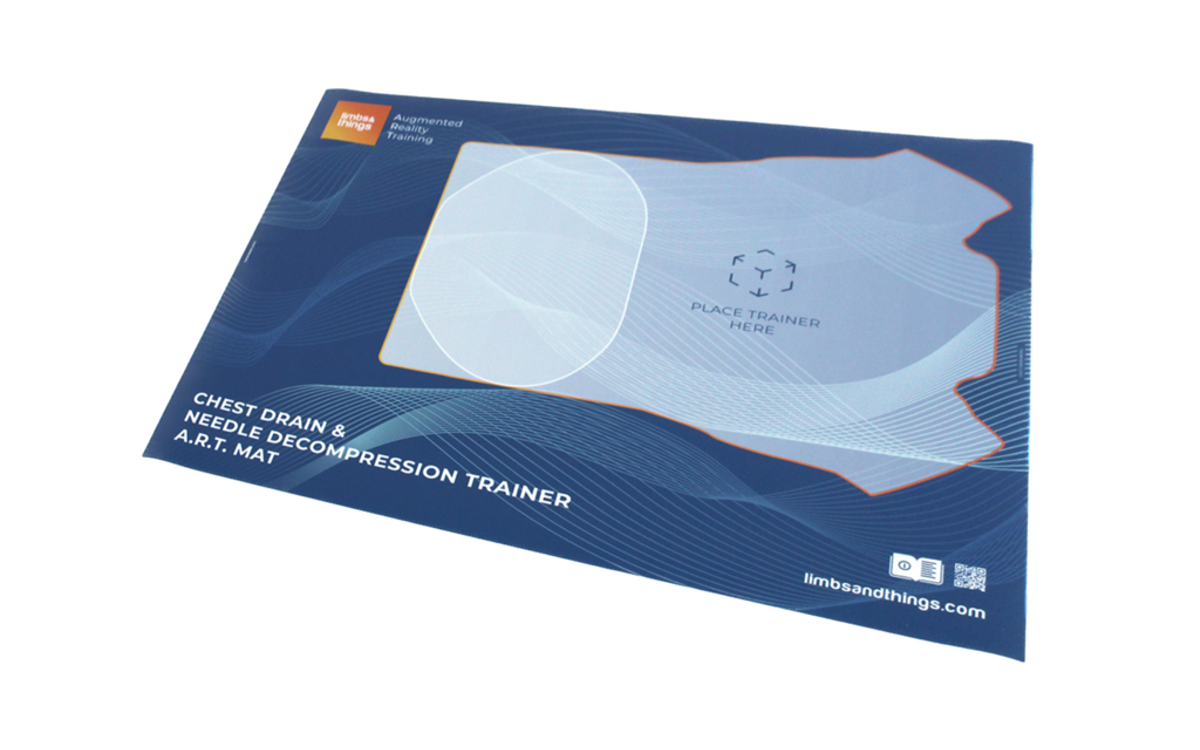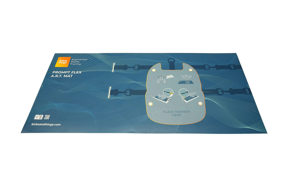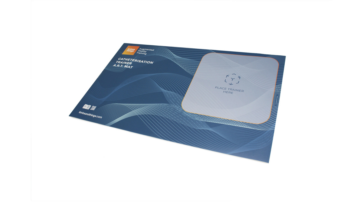
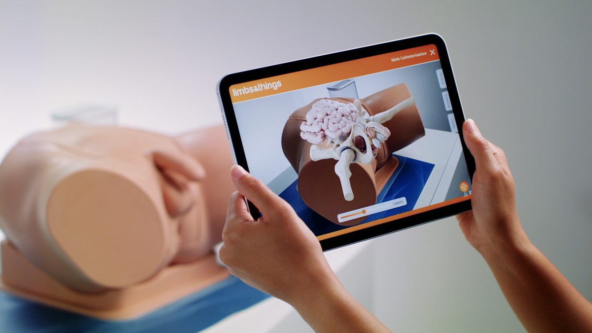
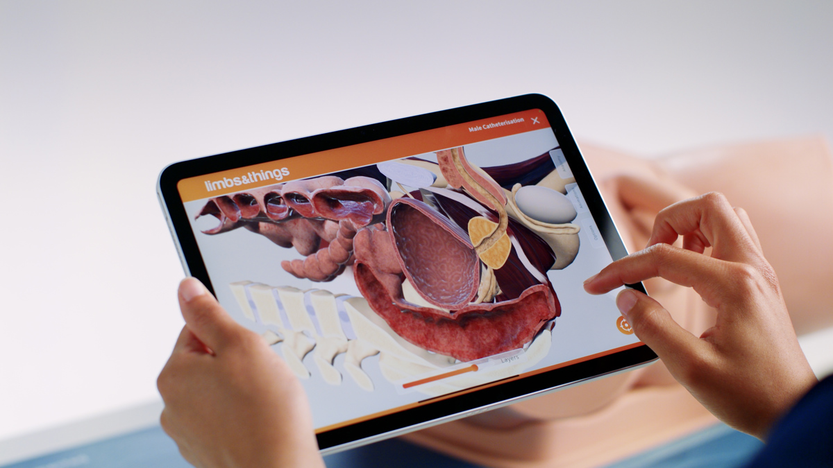

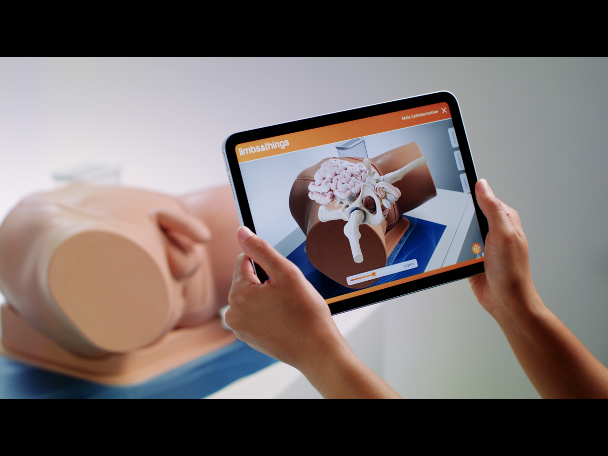

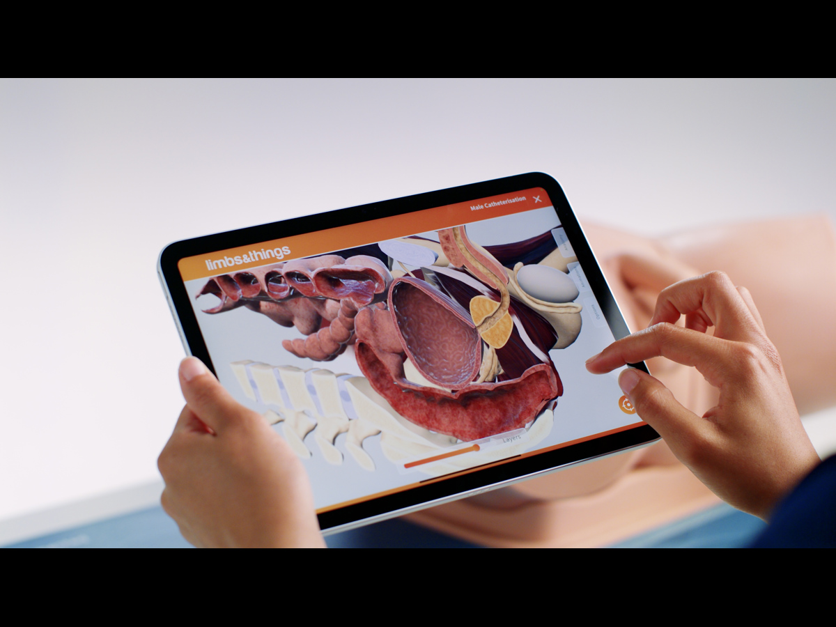
Enhance your existing Catheterisation Trainer learning experience with the latest Augmented Reality Training (ART).
ART Mats are the newest product from Limbs & Things, bringing your Male & Female Catheterisation Trainers to life with the latest AR technology.
Which Catheterisation Trainers work with the new ART Mats?
- Male Catheterisation Trainer Light Skin Tone/60850
- Male Catheterisation Trainer Dark Skin Tone/60870
- Female Catheterisation Trainer Light Skin Tone/60851
- Female Catheterisation Trainer Dark Skin Tone/60869
- Standard Catheterisation Set Light Skin Tone/60853, and Dark Skin Tone/60871
- Advanced Catheterisation Trainer Set Light Skin Tone/60854
Enhance your existing Catheterisation Trainer learning experience with the latest Augmented Reality Training (ART).
ART Mats are the newest product from Limbs & Things, bringing your Male & Female Catheterisation Trainers to life with the latest AR technology.
Which Catheterisation Trainers work with the new ART Mats?
- Male Catheterisation Trainer Light Skin Tone/60850
- Male Catheterisation Trainer Dark Skin Tone/60870
- Female Catheterisation Trainer Light Skin Tone/60851
- Female Catheterisation Trainer Dark Skin Tone/60869
- Standard Catheterisation Set Light Skin Tone/60853, and Dark Skin Tone/60871
- Advanced Catheterisation Trainer Set Light Skin Tone/60854, and Dark Skin Tone 60872
In addition to the hands-on training made accessible with the Limbs & Things simulation models, the ART Mats let students get under the skin for a deeper understanding of the patient’s anatomy.
Featuring realistic 3D models created from actual MRI and CT datasets, medical artists worked in collaboration with digital experts to create the app’s anatomical and skeletal overlays.
What is augmented reality?
Augments Reality (AR) is the combination of computer generated imagery superimposed on real world environments to create an interactive view.
How are Limbs & Things using AR technology to improve medical training?
At Limbs & Things we understand that great medical training gives students a deeper understanding of procedures and the human body.
As such, we’ve combined real world MRI and CT scan data, with the skills of talented medical artists and digital creators, to bring the internal anatomy of our trainers to life.
Within the app’s digital environment, you can move around your task trainer and view various overlays, including: the musculature, organs and vessels, and skeletal structure. The interface allows you to move seamlessly between the layers, as well as view their cross sections.
Students are also able to view digital procedures in the AR environment to see how the procedure is done, and its impact on the patient’s anatomy.
How does the 3D interactive space work?
Even without access to the trainer and mat, students will be able to explore the related anatomy within the app’s interactive space.
The 3D modelling gives you the same, anatomically accurate, rendering, that can be manipulated on screen to reveal the layers of the trainer, and demonstrate procedures with step by step labelling.
*Note: This product does not come with a tablet.
Overview
- Enhances Catheterisation training with an interactive 3D space and augmented reality anatomy
- Augmented reality visualisations of the task trainer anatomy
- 3D physiology to aid understanding of the effect of procedures on the body
Realism
- Anatomically accurate 3D models and illustrations
- Illustrations created by medical artists, from MRI and CT datasets, as well as anatomical atlases and medical research data
Versatility
- Portable for ease of use with the task trainer on any flat surface
- Apps are available for both Android and iOS devices
Cleaning
- The ART Mat can be wiped with a soft damp cloth if needed
- Allow to dry thoroughly before storing
- Ensure device camera is clean, for best performance
Safety
- Always be aware of your surroundings when using the interactive features
- Roll to store, DO NOT fold
- Never move the mat when a task trainer is placed on it
Anatomy
Male Catheterisation anatomy:
- Muscles of the pelvis and pelvic floor
- Structures of the penis, urethra, prostate and bladder
- Variations of these structures that may impact the procedure, such as, urethral and prostatic stricture, and distended bladder
Visualisations of the internal anatomy, including cross sections:
- External skin, in both light and dark skin tones
- Muscles of the pelvis and pelvic floor
- Organs of the pelvis and reproductive system: bladder, vas deferens and urethra, prostate, testicles and structures of the penis
Female Catheterisation anatomy:
- Muscles of the pelvis and pelvic floor
- Structures of the vulva, vagina, urethra, uterus and bladder
- Variation to include potential distended bladder
Visualisations of the internal anatomy, including cross sections:
- External skin, in both light and dark skin tones
- Muscles of the pelvis and pelvic floor
- Organs of the pelvis and reproductive system: uterus and vagina with associated structures, bladder
Skills Gained
- Enhanced understanding of physiology, including geriatric and high BMI patients
- Conceptualisation of procedures
Works with the following products:
-
Light
-
Dark
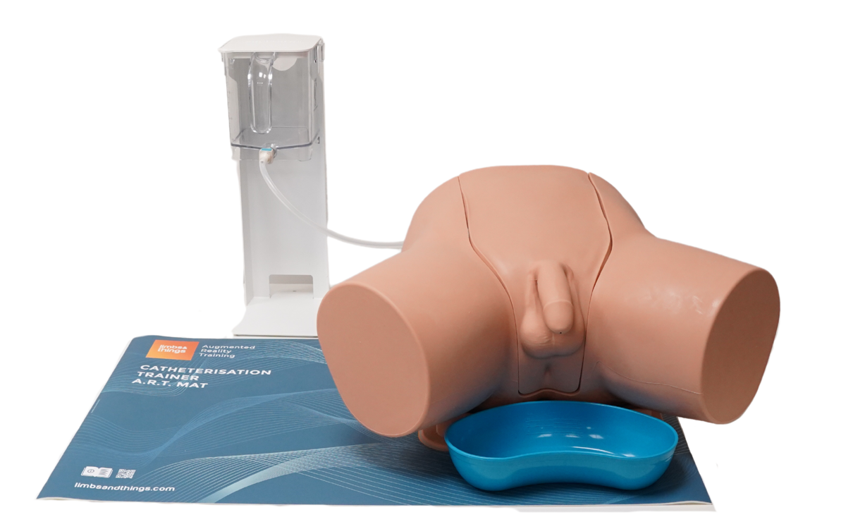
Manlig kateteriseringsattrapp
-
Light
-
Dark
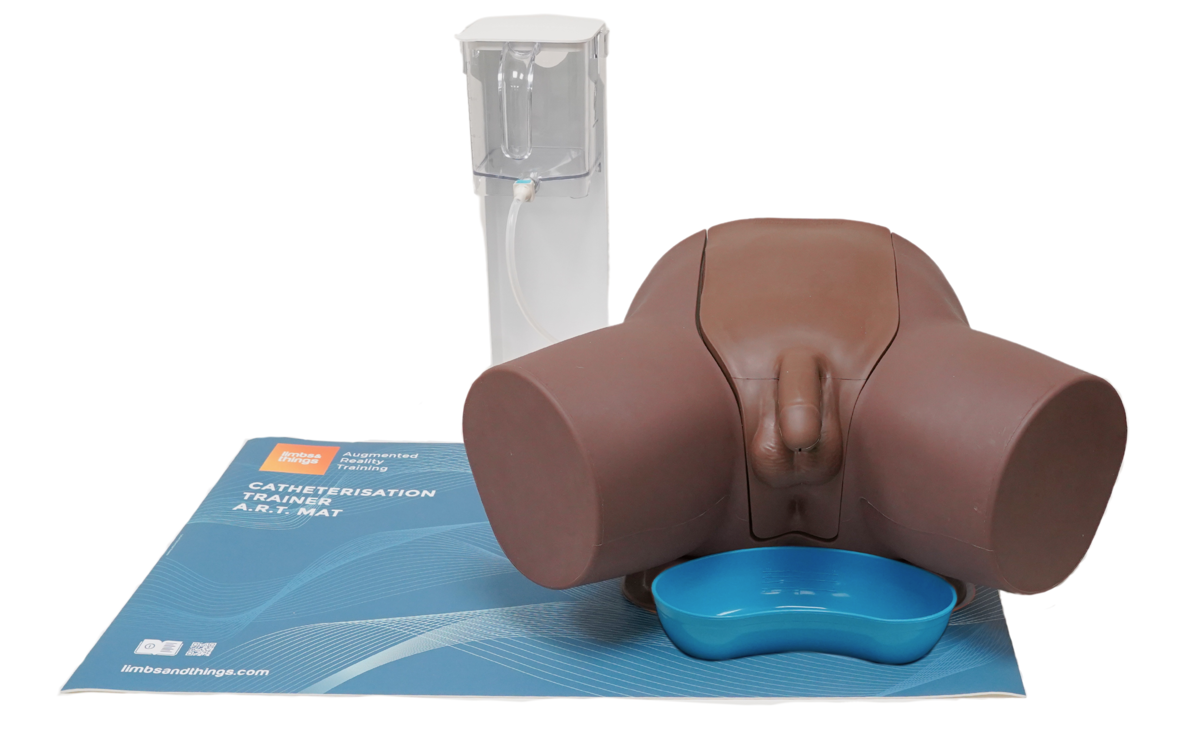
Manlig kateteriseringsattrapp - Mörk
-
Light
-
Dark
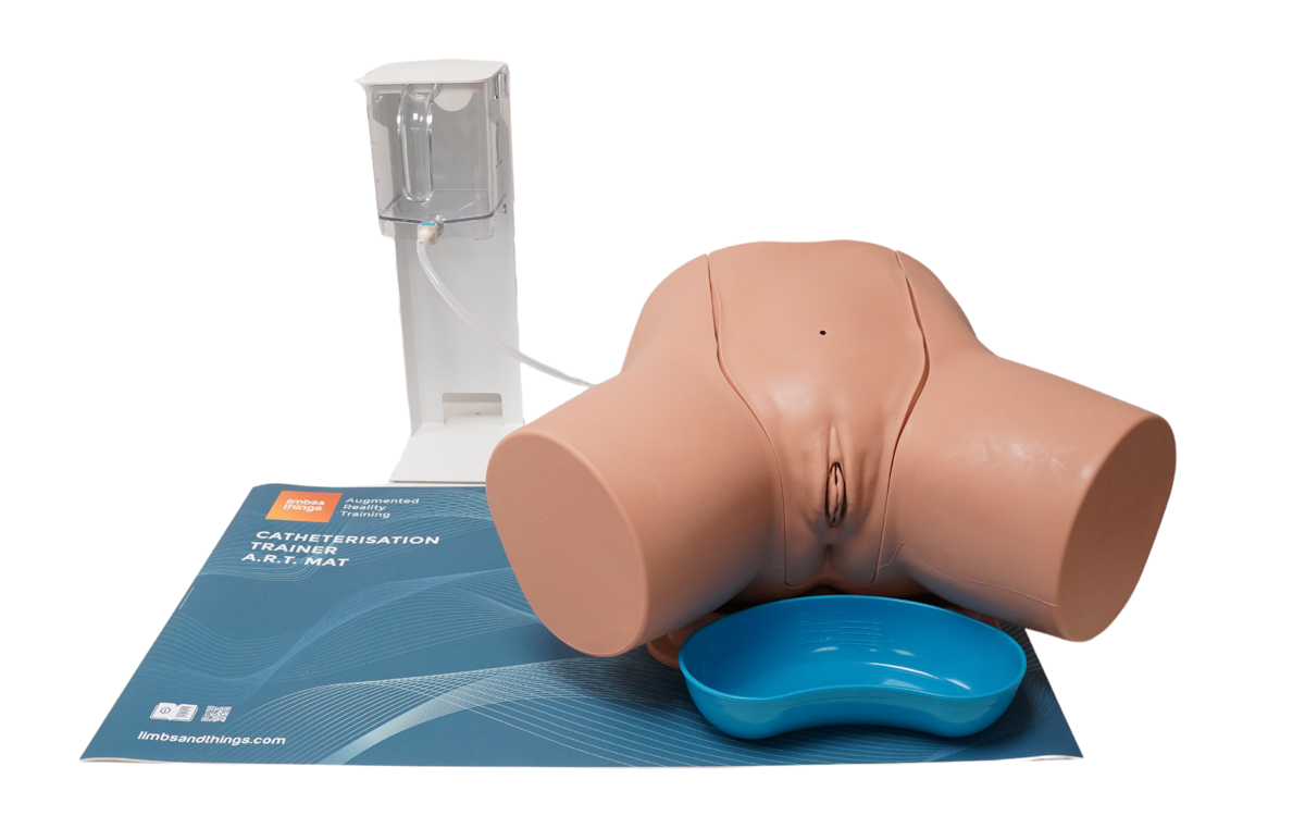
Kvinnlig kateteriseringsattrapp
-
Light
-
Dark
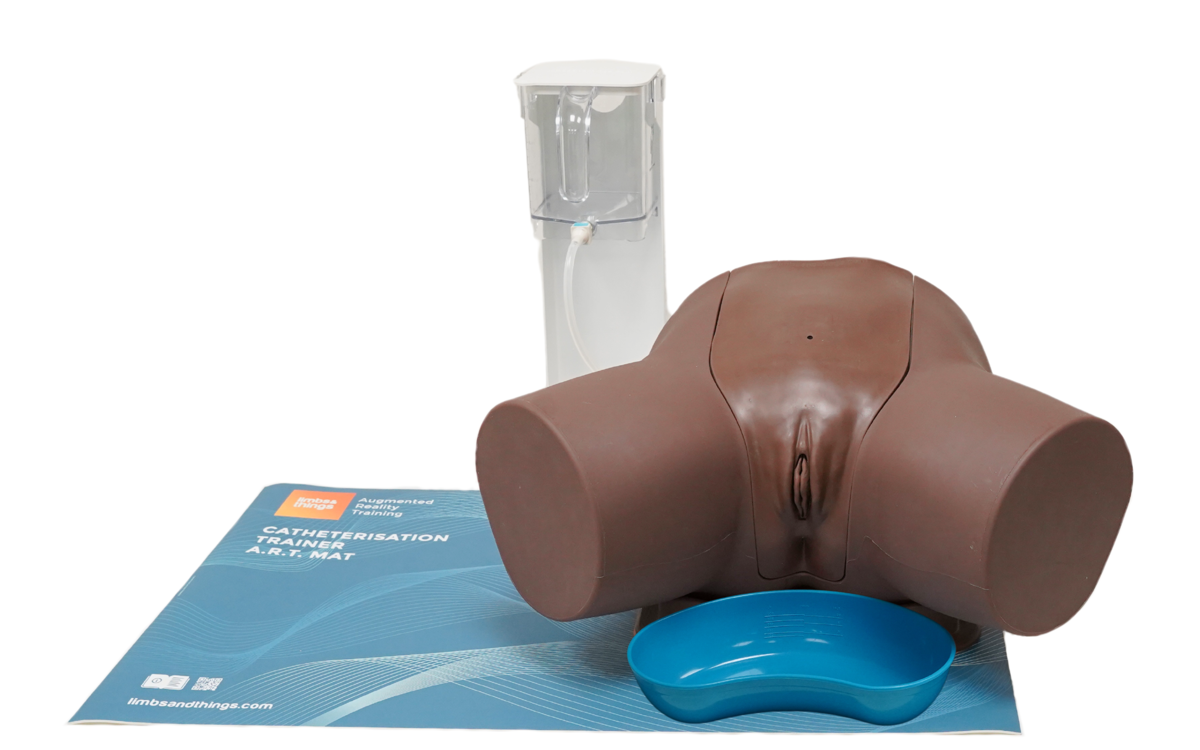
Kvinnlig kateteriseringsattrapp - Mörk
-
Light
-
Dark
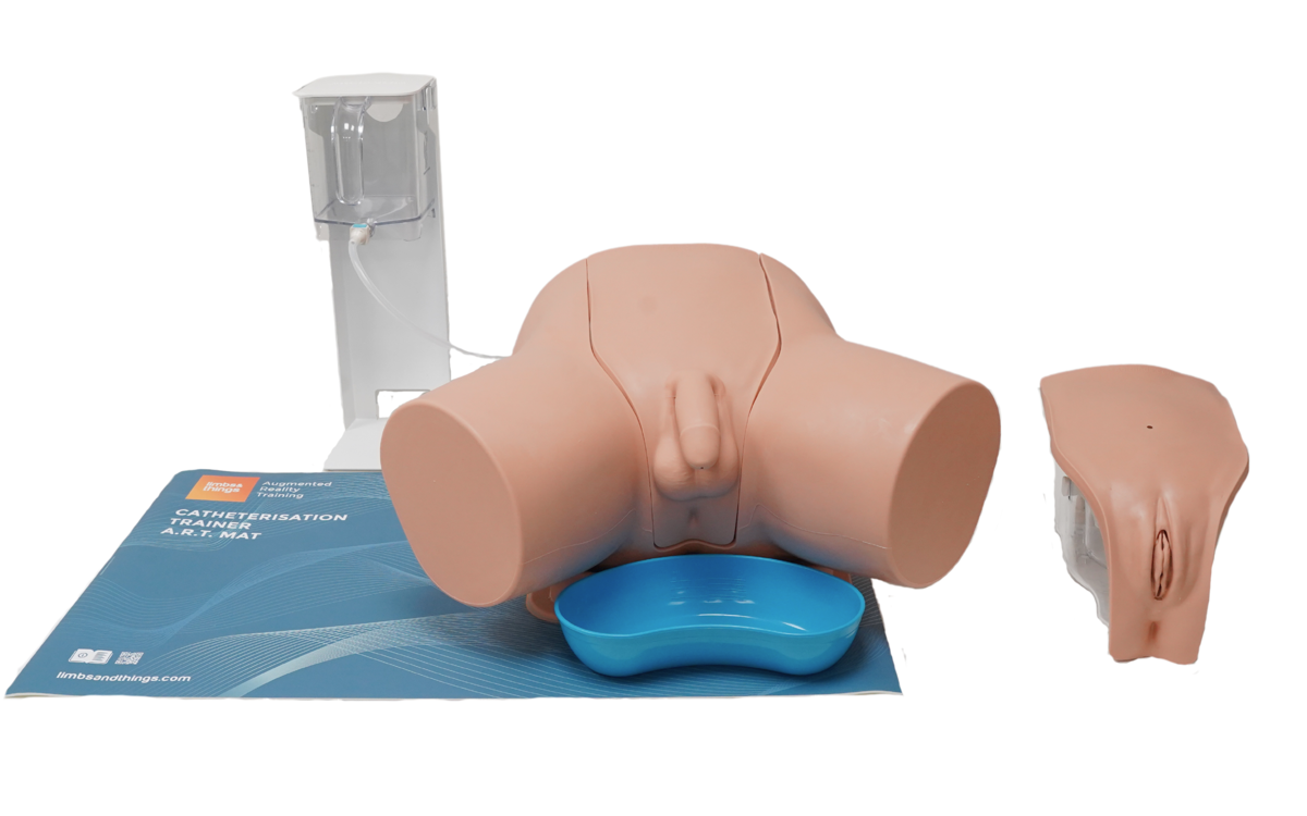
Sats med standardkateteriseringsattrapper
-
Light
-
Dark
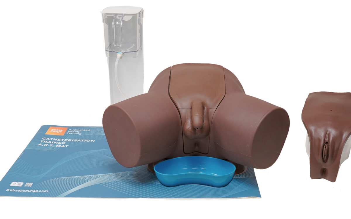
Sats med standardkateteriseringsattrapper - Mörk
-
Light
-
Dark
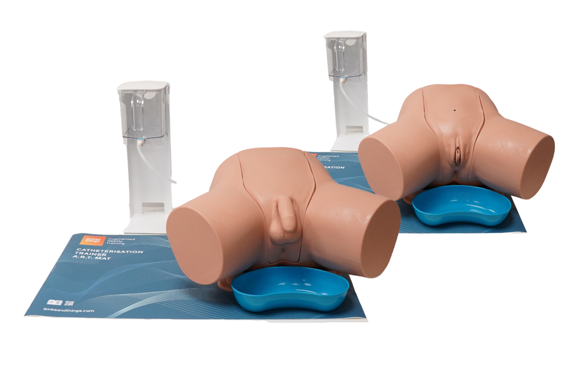
Sats med avancerade kateteriseringsattrapper
-
Light
-
Dark
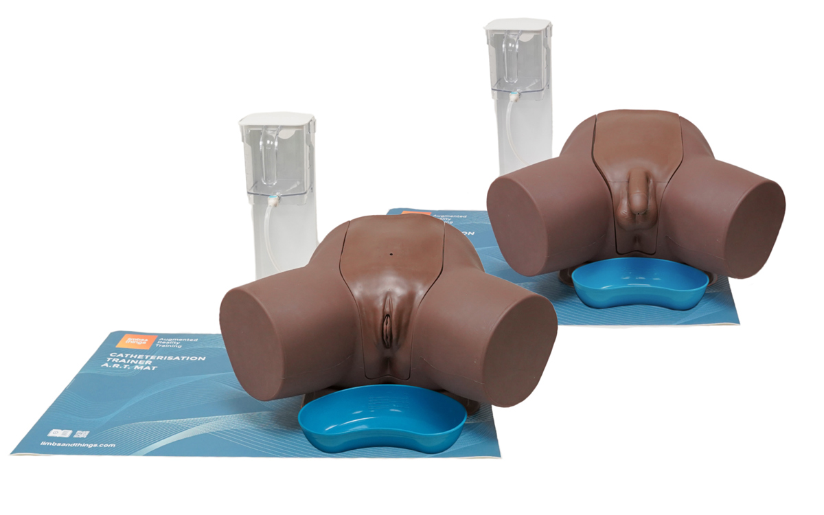
Sats med avancerade kateteriseringsattrapper - Mörk
Device requirements for running the ART App:
iOS Devices:
- Devices that support ARKit
- For specific device information, visit Apple’s Augmented Reality page for compatibility
Android Devices:
- Devices that support ARCore SDK
- For specific device information, visit Google for Developers’ ARCore page for compatibility
The app won’t scan the ART Mat correctly, what do I do?
If the mat is not being recognised by your device camera, we recommend trying the following troubleshooting points:
- Ensure that the mat is on a flat surface in a well-lit place, we would recommend a consistent light source as opposed to natural light
- Remove anything from the surface of the mat that is not the model, check the mat is clean and the pattern is not obstructed by any marks, ensure the trainer is positioned within the designated box
- When scanning the mat with your device’s camera, hold steady for at least 10 seconds
- If the above points do not resolve the issue, fully close the app and restart it
- If problems persist, please contact customer services or your local sales representative
Which Catheterisation Trainers work with the new ART Mats?
You can use the following trainers with the ART Mats:
- Male Catheterisation Trainer Light Skin Tone/60850
- Male Catheterisation Trainer Dark Skin Tone/60870
- Female Catheterisation Trainer Light Skin Tone/60851
- Female Catheterisation Trainer Dark Skin Tone/60869
- Standard Catheterisation Set Light Skin Tone/60853, and Dark Skin Tone/60871
- Advanced Catheterisation Trainer Set Light Skin Tone/60854, and Dark Skin Tone 60872
Note: Suprapubic procedures and anatomy can be found within the Female model section of the app.
I’ve scanned the Catheterisation trainer on the mat, but the digital overlay isn’t in the correct place – what do I do?
You can easily re-scan your task trainer and mat in the app to realign the model and overlay.
Check the ART User Guide for the correct way to position the model, this is within the marked section of the mat. Position your device so that the mat and model are set up clearly in frame, with no obstructions.
For a successful screen capture, we recommend that you hold the tablet as steady as possible with the product within the screen’s boundaries for around 10 seconds, until the augmented reality anatomy diagram appears over your screen image.
Are there specific mobiles devices that we will be able to use the Limbs & Things ART App on?
The Limbs & Things ART App is available to download for free on the App Store or Google Play, and is compatible with iOS and Android systems. Check the Download tab for specifications and links to the app stores with detailed product lists.
Note: Though you can access the features through a smartphone, we would recommend the use of a tablet to get the best experience.
Can we have more than one student studying with the catheterization trainer and ART Mat?
Yes, the AR simulator can be used by multiple trainees at one time.
Each trainee would need a compatible smart device with the ART App download. When you’ve set up the trainer, it can be scanned by devices from multiple angles, allowing them to join sessions at any time.
Once scanned, students will be able to move around the product to examine the anatomy at any angle, however, to get the best view of the cross sections, the user would need to be positioned towards the left hand side of the mat.
Note: The initial scan to connect the AR environment with the trainer requires the device to be able to see the left hand side (“empty” side) of the mat.
How do I turn on the anatomy labels within the virtual environment?
Within the interactive 3D mode, on the right hand side tabs, click options and toggle the labels on.
How can I switch between the different skin tones when viewing the anatomy?
Within the interactive 3D mode, on the right hand side tabs, click options and toggle between light and dark.

